Skip to the content
About
Team Curezone
FAQ
Payment and Insurance
Gallery
Contact Us
Locations
Heartland – Mississauga
Erin Mills – Mississauga
Burloak – Oakville
Services
Pre-Post Surgical Rehabilitation
Laser Therapy
McKenzie Spinal Therapy
Pelvic Health Physiotherapy
Vestibular Rehabilitation
TMJ Physiotherapy
Concussion Treatment
Shock Wave Therapy
Spinal Decompression Therapy
Motor vehicle accident Injury
Balance & Coordination
Chiropody Treatment
Chiropractic Treatment
Compression Stockings
Sports Injury
Workplace Injury
Massage Therapy
Acupuncture Treatment
Blog
Book Now
Home
Blog
Injury
Physiotherapy
April 24, 2024
Physiotherapy for ACL injury
Physiotherapy
April 22, 2024
Physiotherapy for Heel PAIN
Pelvic Floor Physiotherapy
April 12, 2024
Ankylosing Spondylitis Physiotherapy
Arthritis
Physiotherapy
April 8, 2024
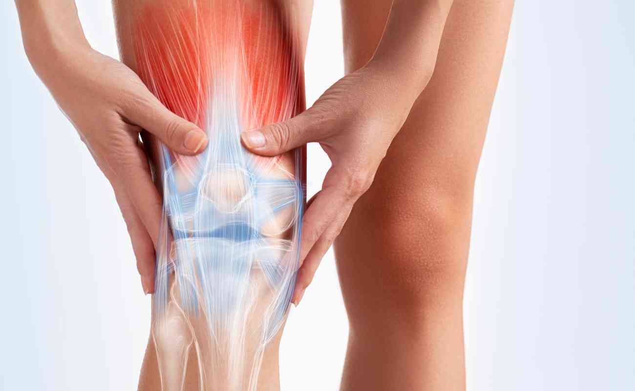
Knee Arthritis Physiotherapy
Physiotherapy
April 5, 2024
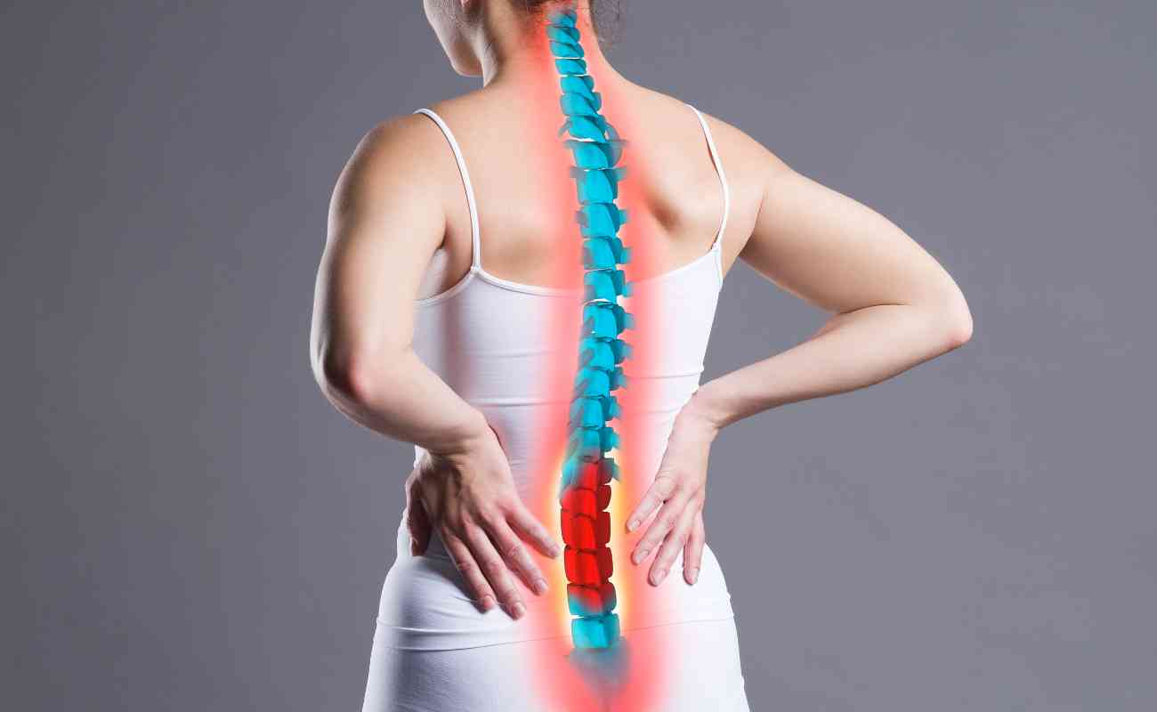
Disc Herniation Physiotherapy
Physiotherapy
April 4, 2024
TMJ Physiotherapy in Mississauga and Oakville
Injury
Physiotherapy
April 3, 2024
Physiotherapy for Boxing Injuries
Injury
Physiotherapy
April 1, 2024
Physiotherapy for Meniscus Injury
Injury
Physiotherapy
March 30, 2024
Physiotherapy for Shoulder Dislocation
Injury
Physiotherapy
March 27, 2024
Physiotherapy for injuries from Kayaking and Rowing
Injury
Physiotherapy
March 25, 2024
Physiotherapy for Martial Arts Injuries
Injury
Physiotherapy
March 22, 2024
Workplace Injury and Physiotherapy (WSIB)
Injury
Physiotherapy
March 17, 2024
Physiotherapy for Skiing Injuries
Arthritis
Physiotherapy
March 14, 2024
Physiotherapy for Osteoporosis
Physiotherapy
March 11, 2024
Carpel Tunnel Syndrome and Physiotherapy
Physiotherapy
Vestibular Physiotherapy
March 8, 2024
Vestibular Rehabilitation Therapy
Injury
Physiotherapy
March 3, 2024
Physiotherapy for Basketball Injuries
Injury
Physiotherapy
Sports Physiotherapy
March 1, 2024
Physiotherapy for Cricket Injuries
01
02
03
…
04
05
06
07
08